Lower Leg Bones Diagram / Anatomy Of Leg And Foot | Anatomi manusia, Anatomi, Biologi - Fibula—thinner, long bone of the lower leg.
byDavid Reynolds•
0
Lower Leg Bones Diagram / Anatomy Of Leg And Foot | Anatomi manusia, Anatomi, Biologi - Fibula—thinner, long bone of the lower leg.. The foot bones shown in this diagram are the talus, navicular, cuneiform, cuboid, metatarsals and calcaneus. The human leg is the entire lower extremity or limb12 of the human body, including the foot, thigh and even the hip or gluteal region; It is situated on the lateral (or little toe) side of the leg. The lower limbs include the bones of the thigh, leg, and foot. The tibia (also called the shinbone) is located these blood vessels supply oxygen and nutrients to the surrounding structures—bones, muscles and nerves.
An intermediate segment, the tibia. When you stand or walk, all the weight of your upper body rests on them. Leg length discrepancy (lld) or anisomelia, is defined as a condition in which the paired lower extremity limbs have a noticeably unequal length. An easy source to get a ton of information easily. The knee joint is the largest joint in the body and is primarily a hinge joint, although some sliding and rotation occur.
Pictures Of Bones Of The Lower Extremities from healthiack.com Continue scrolling to read more below. The bones of the human leg, like those of other mammals, consist of a basal segment, the femur (thighbone); The knee joint is the largest joint in the body and is primarily a hinge joint, although some sliding and rotation occur. The human leg, in the general word sense, is the entire lower limb of the human body, including the foot, thigh and even the hip or gluteal region. The two bones beneath your knee that make up your shin are your tibia and fibula. Vector illustration with human skeleton scheme isolated on a white background. The knee joint is the largest joint in the body and is primarily a hinge joint, although some sliding. Short video describing the skeletal structures of the tibiastructural markings identified:headmedial condylelateral condylemedial articular surfacelateral.
Human anatomy diagrams show internal organs, cells, systems, conditions, symptoms and sickness information and/or tips for healthy living.
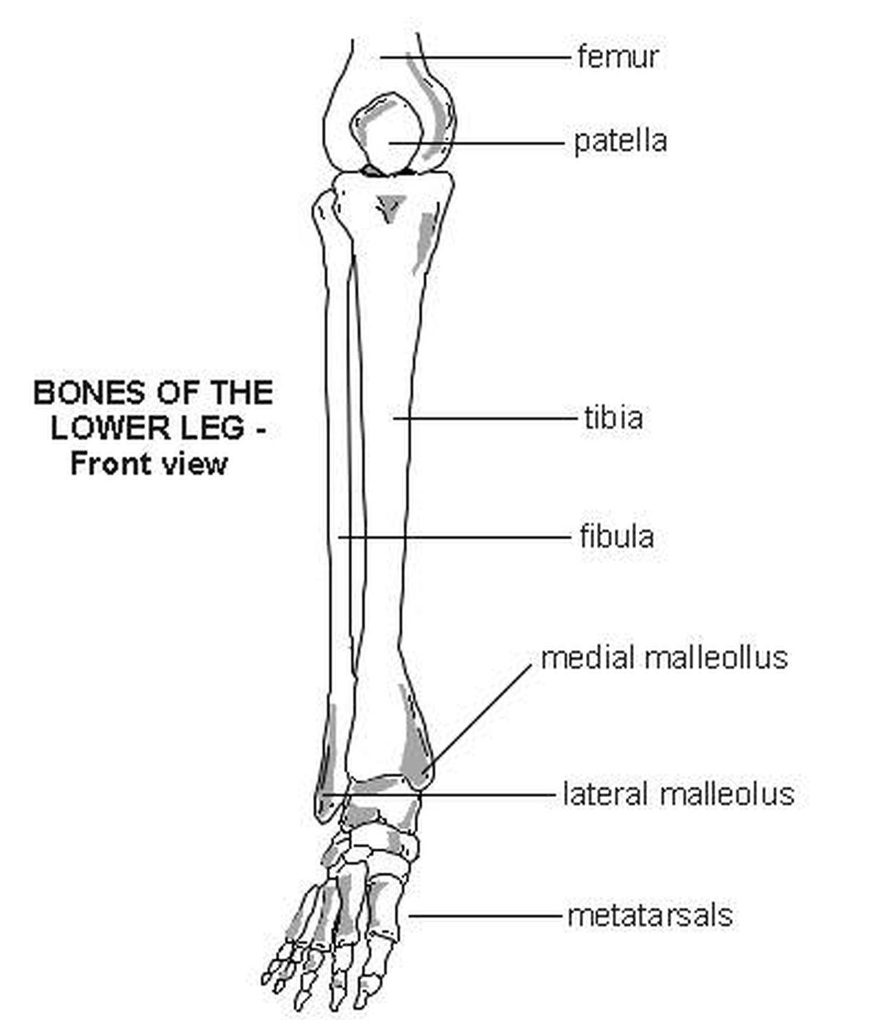
The tibia (shin bone) is the medial bone of the leg and is larger than the fibula, with which it is paired (figure 3). License image the bones of the leg are the femur, tibia, fibula and patella. The tibia (also called the shinbone) is located these blood vessels supply oxygen and nutrients to the surrounding structures—bones, muscles and nerves. We will first just take a general look at the skeleton of the lower limb and then, consider the bones in more detail when we get to each region. An easy source to get a ton of information easily. Hips, shoulders, arms, and legs: The fibula is a long, skinny lower leg bone that looks rather fragile. The two bones beneath your knee that make up your shin are your tibia and fibula. Leg length discrepancy (lld) or anisomelia, is defined as a condition in which the paired lower extremity limbs have a noticeably unequal length. The upper leg bone is connected to the lower leg bones at the knee by a hinge joint. Diagram of lower leg bones posted on march 25, 2019 by admin this image shows the structure of tibia and fibula left panel legs bone diagram 20 13 asyaunited de u2022 hip drawing outline foot overview of bones the lower limb posterior and anterior view respectively 62 infographic diagram of human skeleton. The bones of the leg are the femur, tibia, fibula and patella. Fibula—thinner, long bone of the lower leg.
The foot bones shown in this diagram are the talus, navicular, cuneiform, cuboid, metatarsals and calcaneus. Leg length discrepancy (lld) or anisomelia, is defined as a condition in which the paired lower extremity limbs have a noticeably unequal length. Most bones (particularly the long bones of the arms and legs — which make up the appendicular skeleton) have a hard outer shell known as cortical bone. The bones of the leg are the femur, tibia, fibula and patella. The human leg is the entire lower extremity or limb12 of the human body, including the foot, thigh and even the hip or gluteal region;
Lower Leg Bones | Anatomy bones, Anatomy and physiology ... from i.pinimg.com Leg, limb or appendage of an animal, used to support the body, provide locomotion, and, in modified form, assist in capturing and eating prey (as in spiders and insects). Vector illustration with human skeleton scheme isolated on a white background. However, in the world of anatomy, the 'leg' strictly means the portion. An easy source to get a ton of information easily. At the distal end of the femur, two rounded condyles meet the tibia and fibula bones of the lower leg to form the knee joint. Leg length discrepancy (lld) or anisomelia, is defined as a condition in which the paired lower extremity limbs have a noticeably unequal length. The bones of the human leg, like those of other mammals, consist of a basal segment, the femur (thighbone); Most bones (particularly the long bones of the arms and legs — which make up the appendicular skeleton) have a hard outer shell known as cortical bone.
Anterior view with primary bones names.
Its lower end helps talus : Anterior view with primary bones names. Leg bones, learn what and where these are as well as their functions and how we use them. Continue scrolling to read more below. The lower leg contains two major long bones, the tibia and the fibula, which are both very strong skeletal structures. (2) hip bone attaches legs to our body. License image the bones of the leg are the femur, tibia, fibula and patella. Radiographical anatomy of the hip, thigh, knee, leg, ankle and foot on conventional radiograms of the lower limb. This diagram shows the skeletal structure of the leg (anterior view) and foot (dorsal view). We will first just take a general look at the skeleton of the lower limb and then, consider the bones in more detail when we get to each region. The thigh and leg bones articulate at the knee joint that is protected and enhanced by the patella bone that supports the quadriceps tendon. Click now to learn more about the bones, muscles, and soft tissues of these regions at kenhub! The human leg is the entire lower extremity or limb12 of the human body, including the foot, thigh and even the hip or gluteal region;
An intermediate segment, the tibia. The knee is a strong but flexible hinge joint that uses. At the distal end of the femur, two rounded condyles meet the tibia and fibula bones of the lower leg to form the knee joint. We will first just take a general look at the skeleton of the lower limb and then, consider the bones in more detail when we get to each region. It is situated on the lateral (or little toe) side of the leg.
muscles of the lower leg - Google Search | Athletic ... from i.pinimg.com Continue scrolling to read more below. Most bones (particularly the long bones of the arms and legs — which make up the appendicular skeleton) have a hard outer shell known as cortical bone. The lower leg constitutes a major portion of a. Tarsals—small bones of the hindfoot. The fibula is a long, skinny lower leg bone that looks rather fragile. Your upper and lower leg are connected by a hinge joint. Human anatomy diagrams show internal organs, cells, systems, conditions, symptoms and sickness information and/or tips for healthy living. Fibula—thinner, long bone of the lower leg.
The tarsals are ankle bones and, along with the other bones in the foot (the metatarsals and phalanges), support weight and act as shock absorbers for the body.
Standard radiography view of anatomical structures of the lower limb. The upper leg bone is connected to the lower leg bones at the knee by a hinge joint. The human leg is the entire lower extremity or limb12 of the human body, including the foot, thigh and even the hip or gluteal region; Leg length discrepancy (lld) has been a controversial issue among researchers and clinicians for many years. This diagram shows the skeletal structure of the leg (anterior view) and foot (dorsal view). This bone creates the lower portion of the ankle joint. Continue scrolling to read more below. Vector illustration with human skeleton scheme isolated on a white background. The foot bones shown in this diagram are the talus, navicular, cuneiform, cuboid, metatarsals and calcaneus. Master leg and knee anatomy using our topic page. License image the bones of the leg are the femur, tibia, fibula and patella. The musculoskeletal segment of the leg, including the foot bones (ankle, heel bone, toe bones), fibula and tibia, knee, femur and femoral neck, hip. There is also a knee cap called patella.
Leg length discrepancy (lld) or anisomelia, is defined as a condition in which the paired lower extremity limbs have a noticeably unequal length leg bones diagram. There is also a knee cap called patella.